Exocrine glands histology labeled 333285
View videos describing the histological features of all the exocrine glands eg histology or the pancreas Exocrine Glands Introduction and Learning Overview, A Unicellular Gland, Classification of Glands, Mucous and Serous Secretory Units, Simple Straight Tubular Glands, Simple Coiled Tubular Glands, Simple Acinar Glands, Simple BranchedLab 2 Endocrine Anatomy & Histology Reading LABPAQ Endocrine System pages 1434 Objectives 1 To identify the major endocrine glands and tissues of the body 2 To identify the histology of the major endocrine glands and relate their structure to their function Identify the major endocrine glands and tissues of the body 3Basic anatomy of exocrine glands and lymphatic organs The average exocrine gland contains simple cuboidal epithelia ← that form grapeshaped or tubeshaped structures called acini Liquids secreted by the cells of acini enter a duct, which can be made of a simple cuboidal epithelium or simple columnar epithelium ←
Chapter 15 Page 4 Histologyolm 4 0
Exocrine glands histology labeled
Exocrine glands histology labeled-Endocrine System Histology Pineal Gland Pineal Gland Brain Sand surrounded by Pinealocytes Low Magnification High Magnification Pituitary Gland Anterior Lobe Posterior Lobe Anterior Lobe of Pituitary Gland Posterior Lobe of Pituitary Gland Thyroid Gland and Parathyroid Gland Exocrine glands can be classified into a variety of categories in terms of their structure They can be categorized according to the shape of their secretory unit Secretory units shaped as a tube are referred to as tubular, whereas spherical units are referred to as alveolar or acinar, when the pancreas is involved Exocrine glands can also be comprised of both tubular and alveolar secretory


Skin The Histology Guide
Duodenum histology slide labeled diagram In this part of the article, I will show you the duodenum histology slide labeled diagram again so that you may summarize it well The pictures showed the longitudinal section of a duodenum, where you will find almost every structureThe sweat glands are simple tubular exocrine glands that are found in the superficial hypodermis bordering on the dermis They discharge their contents onto the surface of the skin via coiled secretory ducts (see the diagram opposite)This video "Exocrine Glands" is part of the Lecturio course "Histology" WATCH the complete course on http//lecturio/exocrineglands LEARN ABOUT What is
Anatomy app by Dr Ashwani Kumar http//bitly/ashwaniappSerous, Mucous and Seromucous Glands videos for NEET PG Classifications of Exocrine Glands Based on numbers of cell Unicellular Multicellular Goblet Cell 12 Multicellular Exocrine Glands 1 Based on branching pattern of ducts Simple No Branching Compound Branched 13 Multicellular Exocrine Glands 2 Based on Shape of Secretory End Piece Tubular Alveolar / Acinar Tubulo alveolar 14Serous acini Dense, basophilic, PAS intracytoplasmic secretory granules containing amylase Have central lumen that is rarely visible by H&E Mucinous / mucous acini
Introduction At the histological level the pancreas is made up of compound glands in "bunch of grapes" fashion The pancreas has an exocrine and endocrine component The exocrine compnent is demonstrated above in 3D with acini in "cluster of grapes" formation subtended by a duct Courtesy Ashley Davidoff MD a06 Exocrine glands with ductal and acinar portions Acinar portion is serous, mucinous or mixed Acini are lined by luminal cells, which are enclosed by myoepithelial cells;Keratinization covering of dry, tough material that prevents water from passing non keratinization epithelial remains moist and soft glandular epithelium specialized cells that produce and secrete substances exocrine glands secretes products into ducts that open onto surfaces sweat glands endocrine glands
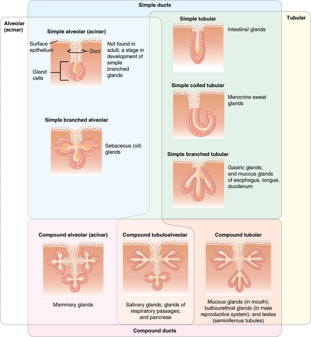


Structure Of Glands Exocrine Endocrine Histology Teachmephysiology
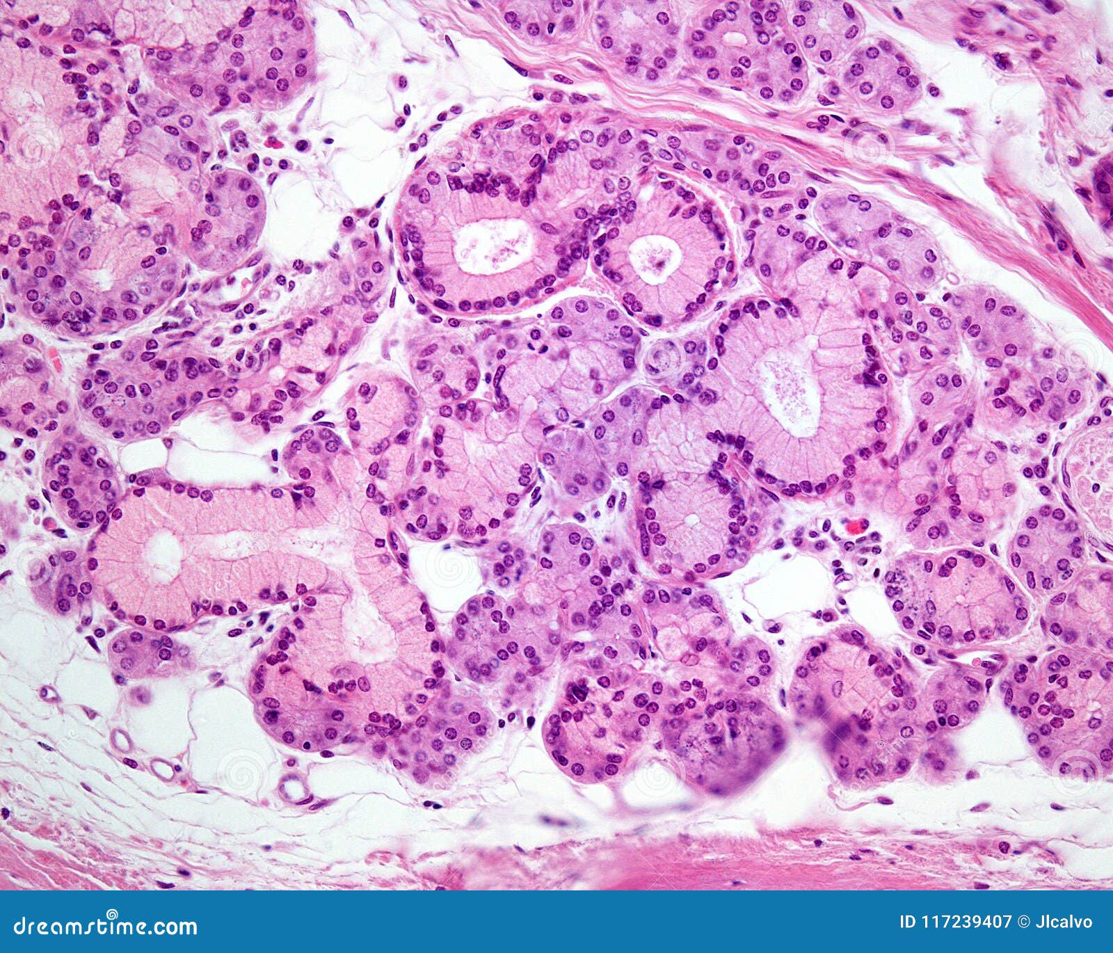


Mixed Exocrine Gland Stock Image Image Of Histology
As in salivary glands, intercalated ductal cells in the pancreas contribute bicarbonate ions (sodium and water follow passively) to the exocrine secretory product However, unlike salivary glands, there are no striated ducts in the pancreas to recover sodium, so the final product is rich in both sodium and bicarbonate (as opposed to saliva inDescribe the histology of the exocrine glands of the Pancreas The exocrine glands of the Pancreas are composed of 3 different cell types and each has a specific function 1 Acinar Cells These cells secrete Zymogen Granules full of digestive enzymes when stimulated and are arranged inComparisons of current secretion and transport models in the mammary gland, exocrine pancreas and salivary gland indicate that significant differences exist between the mammary gland and other exocrine organs in how proteins and lipids are packaged and secreted, and how fluid is transported Mammary Glands, Human / anatomy & histology
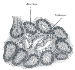


Acinus Wikipedia


Skin The Histology Guide
4 rows The exocrine component of the pancreas makes up about 98% of the pancreatic tissue It isCovers body surface, lines body cavities, lines external/internal organs avascular ( no blood vessels) Functions of Epithelial Tissue protection, secretion, excretion, absorption, filtration, sensation basement membrane anchors an epithelium to the connective tissues Simple Epithelia only one layer of cellsFigure 51 Basic anatomy of an exocrine gland Basic anatomy of exocrine glands and lymphatic organs The average exocrine gland is composed of simple cuboidal epithelial cells that form grapeshaped structures called acini (although other glands have tubeshaped ends) These grapeshaped structures are where secretions are produced


Lab 6 Urinary Exocrine And Endocrine Histology


Tubular Gland An Overview Sciencedirect Topics
A comprehensive account of the anatomy and histophysiology of the ecdysial glands of Philosamia ricini during post embryonic development has been given The study clearly indicates an increase in number of cells in each larval instar by endomitosis The maximum number of Please, try to identify the following histology slides of exocrine and endocrine glands – #1 Parotid salivary gland histology slide #2 Submandibular salivary gland slide #3 Sublingual gland histology slide #4 Mammary gland histology slide #5 Pituitary histology slide #6 Thyroid gland histology slide #7 Adrenal gland microscope slide


Electron Micrograph Of Part Of A Serous Acinus In A Parotid Gland


Biology 2404 Tissues
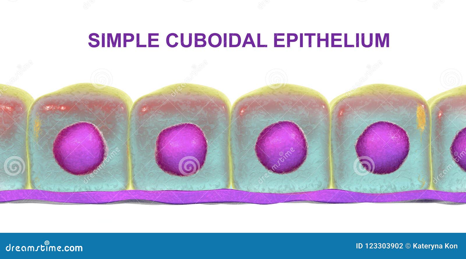


Simple Cuboidal Epithelium Stock Illustration Illustration Of Cell


Blue Histology Epithelia And Glands


Submandibular Gland


Pancreas Histology Identifying Features With Labeled Slide Images Anatomylearner The Place To Learn Veterinary Anatomy Online


Skin The Histology Guide
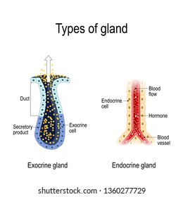


Histology Endocrine Glands Images Stock Photos Vectors Shutterstock
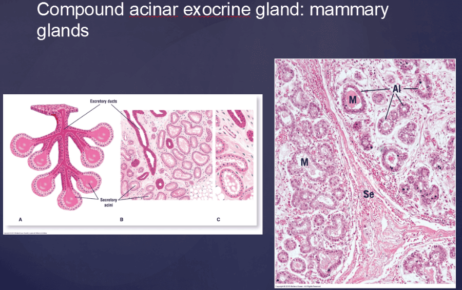


Print Vertibrate Histology Test 1 Flashcards Easy Notecards


Blue Histology Epithelia And Glands
:background_color(FFFFFF):format(jpeg)/images/library/2471/shYLtWdYUjK3qRvwMzK4Q_Compound_tuboalveolar_mixed_salicary_gland.png)


Glands Anatomy And Clinical Notes Kenhub
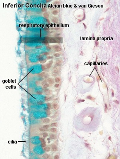


Anat2241 Glandular Epithelia Embryology


Exocrine Glands Of The Integumentary System Youtube


Chapter 15 Page 4 Histologyolm 4 0
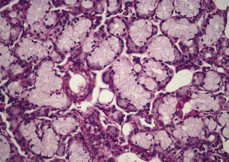


Pathology Outlines Anatomy Histology


Blue Histology Epithelia And Glands
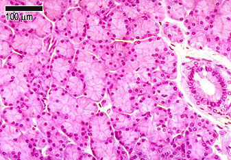


Glandular Tissue The Histology Guide


Sweat Gland Wikipedia


Skin The Histology Guide
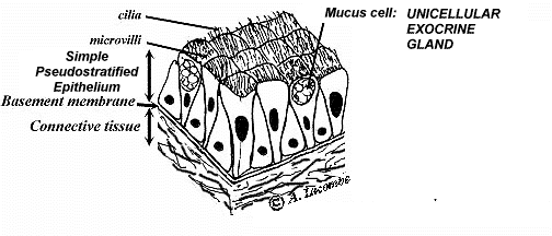


Al S Tutorial Histology Epithelium Glandular Epithelia


Exocrine Pancreas Google Search Pancreas Histology Slides Endocrine System


Salivary Glands Intechopen


Study Notes
:background_color(FFFFFF):format(jpeg)/images/library/2472/r3yQf6wxyufP1joSAhl7eg_Mixed_seromucous_glands.png)


Glands Anatomy And Clinical Notes Kenhub


Picture Test In Histology Of The Endocrine Glands Youtube


Skin Lab
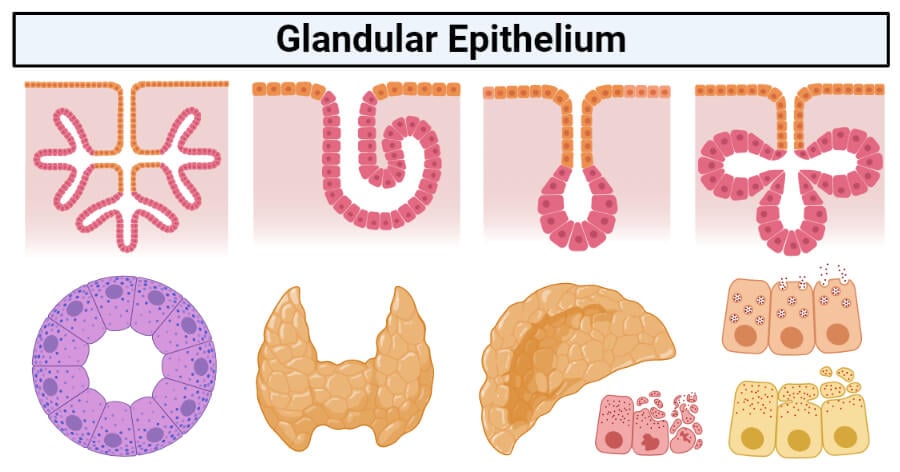


Glandular Epithelium Definition Structure Functions Examples
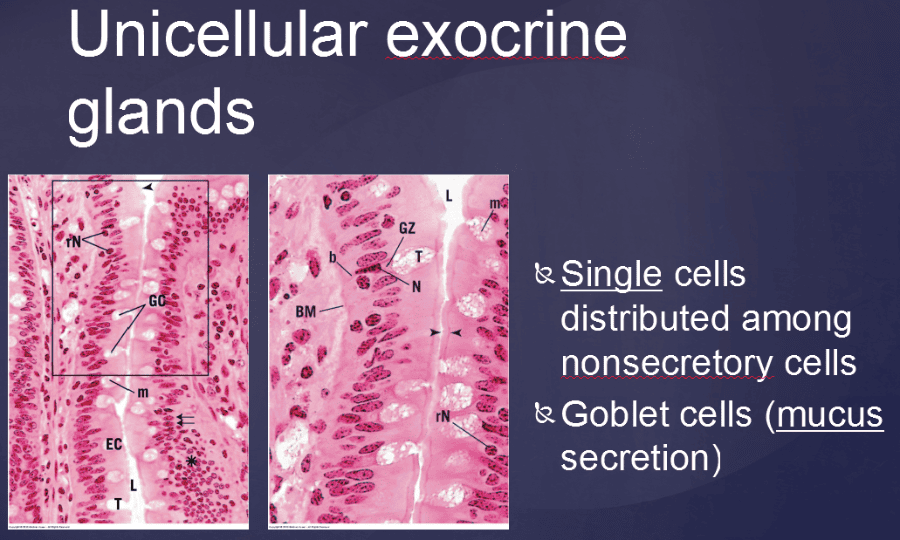


Print Vertibrate Histology Test 1 Flashcards Easy Notecards


Glandular Epithelium And Glands Springerlink


Lab 6 Urinary Exocrine And Endocrine Histology
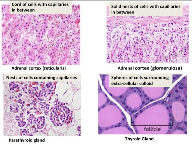


Glands Histology
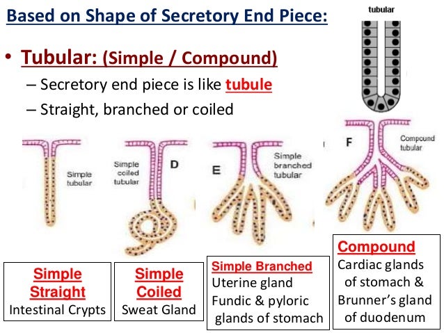


Glands Histology


Animal Tissues Epithelium Salivary Gland Atlas Of Plant And Animal Histology


Salivary Gland An Overview Sciencedirect Topics


Blue Histology Epithelia And Glands


Sweat Glands Preview Histology Function Human Anatomy Kenhub Youtube


Parotid Gland


Pancreatic Histology Exocrine Tissue


Salivary Gland An Overview Sciencedirect Topics


Morphology Of Salivary And Lacrimal Glands Intechopen
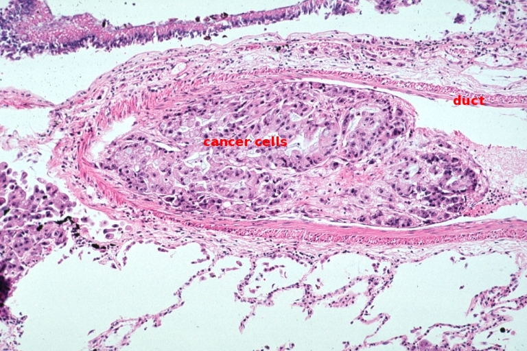


Glandular Epithelium


Morphology Of Salivary And Lacrimal Glands Intechopen


Study Notes


Pin On Histology Slides


Sebaceous Glands An Overview Sciencedirect Topics


Pancreatic Histology Exocrine Tissue
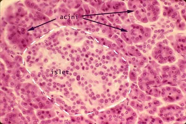


Histology At Siu


Mammary Gland Histology Lactating And Non Lactating Histological Characteristics Anatomylearner The Place To Learn Veterinary Anatomy Online


Pancreas Histology Pancreas Labels Histology Slide Medical School Studying Histology Slides Medical Education
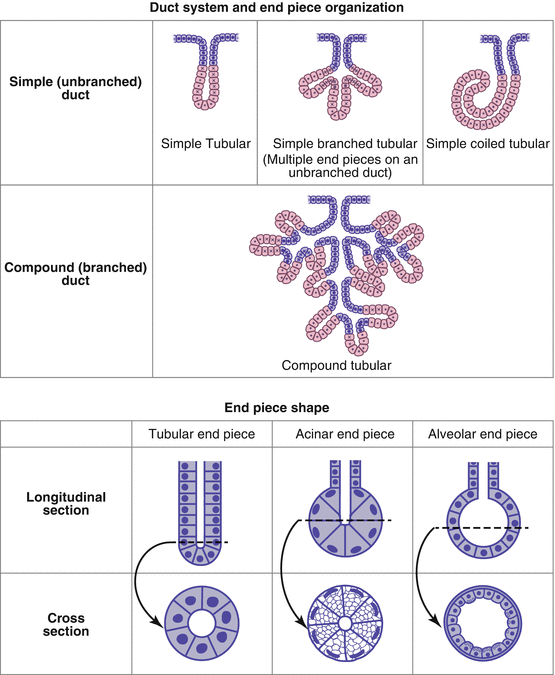


Glandular Epithelium And Glands Springerlink


Chapter 15 Page 5 Histologyolm 4 0


Parotid Gland


Tubular Gland An Overview Sciencedirect Topics
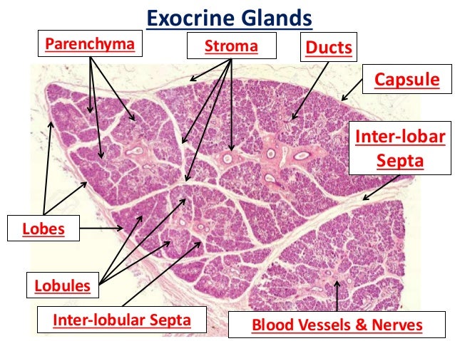


Glands Histology


Endocrine


Light Micrograph Of A Resting Mammary Gland At Higher Magnification


Salivary Gland An Overview Sciencedirect Topics


Animal Tissues Epithelial Tissue Glands Atlas Of Plant And Animal Histology


Lacrimal Gland Histopathology H E Staining Of A Normal Lacrimal Gland Download Scientific Diagram


Intercalated Duct An Overview Sciencedirect Topics


Chapter 15 Page 7 Histologyolm 4 0


Exocrine Gland Wikipedia


Tubular Gland Wikipedia


Microscope Slides Of Cells And Tissues Histology Guide


Epithelial Tissues Exocrine Glands Quiz Digital Histology


Basic Histology Short Columnar Epithelium Exocrine Glands
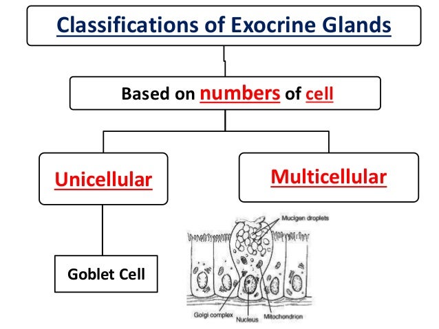


Glands Histology
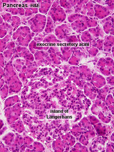


Gastrointestinal Tract Pancreas Histology Embryology
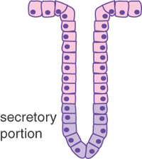


Epithelia The Histology Guide


Pin By Dany Levy On Biology Medicine Histology Slides Medical Coding Pancreas


Chapter 15 Page 4 Histologyolm 4 0


Holocrine Wikipedia
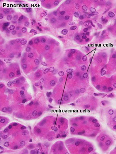


File Pancreas Histology 002 Jpg Embryology


Histology Of Pancreas Youtube
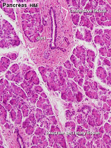


Gastrointestinal Tract Pancreas Histology Embryology


Animal Tissues Epithelial Tissue Glands Atlas Of Plant And Animal Histology
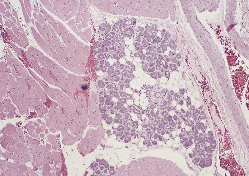


Pathology Outlines Anatomy Histology


Histology Of The Pancreas Endocrine And Exocrine Youtube


Epithelial Glands Basicmedical Key


Lab 6 Urinary Exocrine And Endocrine Histology


Lab 6 Urinary Exocrine And Endocrine Histology


Kgmu Org


Liver And Pancreas


Epithelia The Histology Guide


Oral The Histology Guide
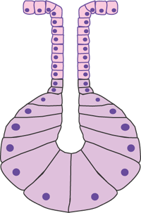


Epithelia The Histology Guide


1


Pancreatic Histology Exocrine Tissue


Salivary Gland Histology Parotid Submandibular And Sublingual Glands Structure Anatomylearner The Place To Learn Veterinary Anatomy Online
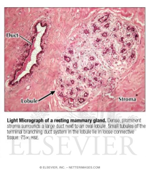


Light Micrograph Of A Resting Mammary Gland


Kgmu Org


Blue Histology Epithelia And Glands


Exercise 4 Epithelium


General Histology Knowledge Amboss


Submandibular Gland
:background_color(FFFFFF):format(jpeg)/images/library/3778/mNlBYB0eHlYXptgna1cnQ_Sweat_glands.png)


Sweat Glands Structure And Function Kenhub


Chapter 15 Page 7 Histologyolm 4 0
:watermark(/images/watermark_5000_10percent.png,0,0,0):watermark(/images/logo_url.png,-10,-10,0):format(jpeg)/images/overview_image/1881/CPsIC67xuz6pLt6JhAM6Wg_pancreas-histology_english.jpg)


Pancreas Histology Exocrine Endocrine Parts Function Kenhub
コメント
コメントを投稿